Posted on

AE: Mammalian Neuronal Monolayers Undergoing Electro Field Stimulation

AE: Mammalian Neuronal Monolayers Undergoing Electro Field Stimulation

I need to do fluorescence microscopy of mammalian, neuronal monolayers that are undergoing electro field stimulation at physiological temperatures in a nourishing environment. Is there a way to create the conditions to conduct this experiment while having direct access to the specimen?

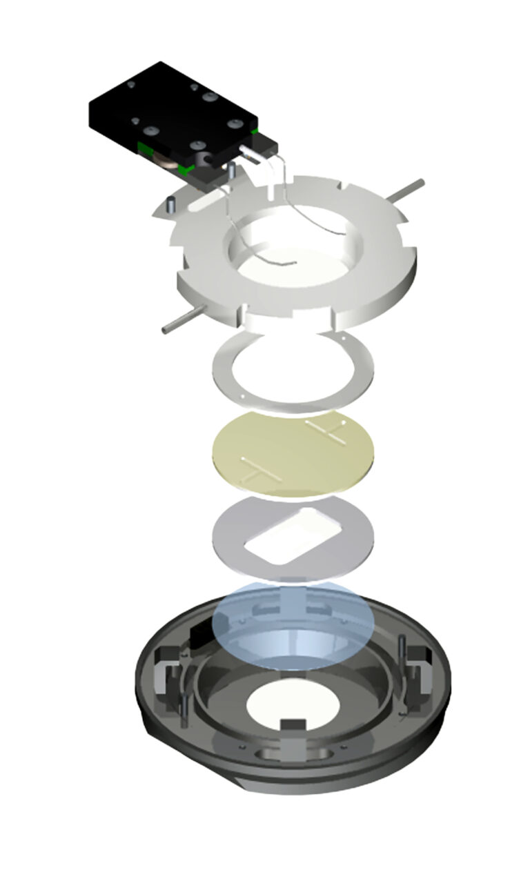
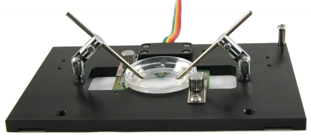
 Let’s look at the physical constraints of your experiment. Since you need direct physical access to your specimen you will need an open dish system.  You will want the highest resolution images you can get so the specimens should be on a glass #1.5 coverslip.  The most efficient means to maintain temperature is direct heating of the coverslip where the cells are adhered.  Note: you don’t want to heat the stage and should only provide heat to the specimen.  In this case your best choice is the Delta T Culture Dish System. It is a coverslip bottomed, open dish micro-environment that heats only the media and specimens in the dish with a very fast, closed loop feedback system to ensure precision temperature control.  This system is designed to be used with numerous accessories that customize it to conform to your experimental constraints.  The following configuration would be ideal for your experiment:  Cells can be plated in a Delta T Dish and have an electrical field introduced to them by a fixture containing bipolar platinum electrodes that are attached to a flip in / flip out support mechanism mounted to a dish retainer.  A supplemental Hinged Perfusion Adapter is used to support perfusion needles for irrigating and aspirating the dish to facilitate long term-experiments. The hinged accessories allow you to easily exchange dishes, maintain registration and re-establish your desired conditions.
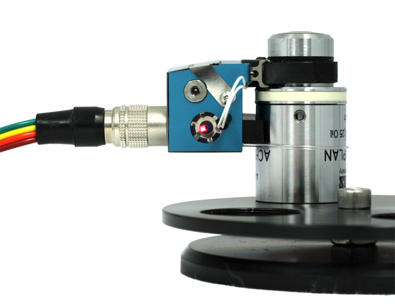
If you are imaging with a high numeric aperture objective, which requires an optical coupling medium, you will also need to warm the objective to prevent it from acting as a thermal sink to your specimen.  When warming an objective it is critical to apply heat to the most efficient thermally receptive location on the objective and incorporate the objective as part of the control loop. These characteristics are exclusive to the Bioptechs Objective Heater to prevent overshoot and establish accurate control.  To reliably and continually perfuse the dish the Delta T micro-perfusion pump is highly recommended.  It will deliver media to the dish at a rate slower than it is removed so the dish cannot overflow. You simply adjust the height of the pickup needle to the level you want in the dish.  This simple configuration is easy to set up, easy to maintain and will offer years of reliable data collection. 

Posted on

Induced change microscopy of live cells

Induced Change Microscopy of Live Cells

My PI just informed me our new project is to investigate the instantaneous effect of a compound on live cells. Some of the setup is intuitive but now I have run into issues I didn’t realize would be problematic. For instance in order to observe my cells what kind of containment is best to use? How do I expose the cells to an alternate media without compromising them? How do I precondition my media? How long does it take to acquire the expected changes? Who can help?

Fortunately, you are not the first person to encounter these issues. As it turns out, this is a routine process used to investigate many cellular reactions. One by one in order, the type of containment vessel should be a parallel plate flow cell having a coverslip surface for imaging. There are several commercial ones available. The one most recommended is the Bioptechs FCS2.


When comparing to the others, the FCS2 was the only one that allows you to select or create any flow geometry you want including laminar flow instead of being rigidly confined to the manufacturers design. It also provided temperature support uniformly over the entire aperture of the chamber. It is compatible with all modes of microscopy you may intend to use and is easy to assemble. When it comes to perfusion you may be thinking it is a plumber’s nightmare but in fact it is already worked out. If you are working with two sources of media, one is just a nutrient media and the other has the variant factor. The plan is to image cells on the scope for about 30 minutes with nutrient media to demonstrate they are “happy” and not compromised in their foreign environment. Then, depending on experimental protocol, introduce the alternate media containing your variant factor while acquiring images of the effects.

If the expected reaction takes place slowly you can manually introduce the variant, but if the protocol requires a rapid introduction of the variant that is where it gets a little tricky and is best to automate the process.  There are two sources of media coming together external to the optical cavity where the cells are plated. Therefore, there is a dead-volume between the adjoining flows and the cells, and a small diffusion gradient that occurs when one flow follows another. The goal is to get the variant factor to the cells at its source concentration ASAP to record the immediate reaction. 

To accomplish this therecomputer programs that control the flow rate of two independent, smooth flow, peristaltic perfusion pumps.

The program allows you to; establish a maintenance flow of nutrient media for a predetermined amount of time that you control, then ramp down the nutrient flow while accelerating the flow of the variant factor, then sustain that rate for a for another determined period of time you chose to ensure the concentration of the variant media is at its peak. The Variant flow rate is then de-accelerated to a minimum rate for imaging. The process can then repeated to rinse the variant factor and restore the prior environmental conditions. This saves a lot of headache
making these transitions especially when it has to be consistently repeated many times for statistical reasons

If you can’t get away with hepes media, you will need to sustain a CO2 tension in the media to maintain proper pH. A number of CO2 systems are available but the one that makes the most sense starts with a 5% CO2 and air tank under pressure then reduces the pressure to ambient with a regulator. That way the gas mixture can be metered out by virtue of its volume with a simple peristaltic pump. If you place a 14 gauge tube into a flask containing media each bubble that rises is about 15 micro-liter. Therefore, all you have to do to establish the flow rate is count the bubbles per minute and do a little arithmetic!

The amount of time it takes to acquire results will depend on the concentration of the variant factor and your cells.

Posted on

Speeding up Cell Plating for Imaging

Speeding Up Cell Plating for Imaging

Sample preparation has always been a notoriously time consuming task that tends to detract from the more essential functions of collecting and analyzing data.  A few key factors come in to play with improving the process of plating cells for imaging and the time that it takes… Cell adhesion, media re-equilibration,  and an unobstructed path for free migration of cells are some of the important factors to improve plating efficiency. 


Cell adhesion is dependent on the surface the cells are being plated on. The chemical composition of the glass affects cell adhesion and all glass surfaces are not created equal.  It is best to use glass that is alkaline free and designed for cell adhesion (check out the Bioptechs Delta T Culture DishesFCS2 coverslips30mm ICD coverslips, and Microaquaduct slides). Sometimes an ECM is required depending on the cell type and protocol, however, in all cases cell plating is improved with the use of Culture Cylinders


A unique attribute of using a Culture Cylinder for plating is the rapid, organic adhesion in a defined location on the dish.  This ensures the best cells are in the most viewable position on your substrate for imaging.  Culture Cylinders have made innovative improvements over pouring tripsinized cells into an entire dish of media and waiting.   Loosely related to cloning rings; Culture Cylinders in contrast are autoclaveable borosilicate glass polished on one surface optically flat to create a hydrostatic seal to the substrate that cells are plated on so that grease is not required.  This minimizes the volume of media used for cells to re-equilibrate to once tripsinized, thereby allowing cells to plate faster.  Also by eliminating the use of grease there is no contamination induced by plating cells or obstructive surface to inhibit cell migration.  As noted in a manuscript by S. Mathupala and A. Sloan (2009) using grease with cloning rings has its own set of inherent problems that have a clear effect on the plating process.  By applying this method, cell plating has become more efficient and also enables a variety of new experimental configurations with multiple Culture Cylinders

Mathupala, S. P., & Sloan, A. E. (2009, April). An agarose-based cloning-ring anchoring method for isolation of viable cell clones. Retrieved August 19, 2016, from http://www.ncbi.nlm.nih.gov/pmc/articles/PMC2727865/